Artwork
Specimen of the embryological head
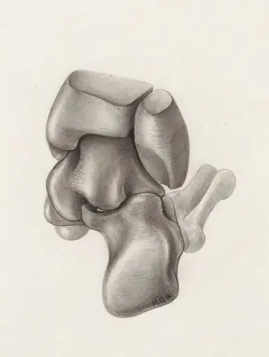
Specimen of the embryological head is a drawing by Unknown. It dates from 1968 and is held in the collection of the Leiden University Libraries. This image is a detailed anatomical drawing depicting the embryonic development of the human head and upper torso.
About this work
Overview
Rendered with delicate shading and minimal linear definition, it captures early fetal form rather than a fully developed human figure.
This image is a detailed anatomical drawing depicting the embryonic development of the human head and upper torso. Rendered with delicate shading and minimal linear definition, it captures early fetal form rather than a fully developed human figure. The absence of sharp contours and the smooth, rounded modeling suggest an intent to illustrate biological growth stages, likely for educational or scientific purposes.
Subject & Meaning
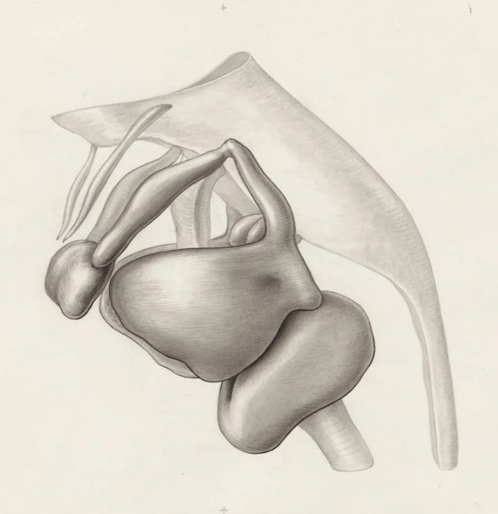
The subject is an early human embryo, shown in a contemplative pose with hands crossed before the face. This positioning may symbolize self-containment or introspection inherent in developmental biology. The distorted proportions—thick neck, abbreviated fingers—reflect embryonic anatomy before ossification and elongation occur, emphasizing the transition from formless mass to structured organism.
Technique & Style
The artist employed soft, graded tonal transitions to suggest volume without relying on outlines, except where the hands meet the face. This approach avoids the rigidity of traditional draftsmanship, favoring a fluid, almost atmospheric rendering. The lack of cross-hatching or stippling distinguishes it from contemporaneous anatomical studies, pointing toward a more observational, less schematic method.
History & Provenance
The work appears to originate from a 19th-century anatomical or embryological study, possibly linked to medical instruction or private research. Its unattributed status and informal presentation suggest it was not intended for public display but rather as a working sketch within a scholarly context, perhaps part of a larger collection of developmental studies.
Context
During the 1800s, advances in microscopy and dissection fueled interest in embryology as a scientific discipline. Drawings like this served as visual records of developmental stages, bridging art and science. Such images were often used in lectures or textbooks to convey complex biological processes to students unfamiliar with live specimens or preserved embryos.
Legacy
Though not widely published or attributed, this drawing exemplifies the quiet intersection of artistic skill and scientific inquiry in pre-photographic anatomy. Its sensitivity to form and subtle modeling influenced later pedagogical illustrations, reinforcing the value of hand-drawn observation in medical education before the dominance of photographic documentation.
Artist & collection
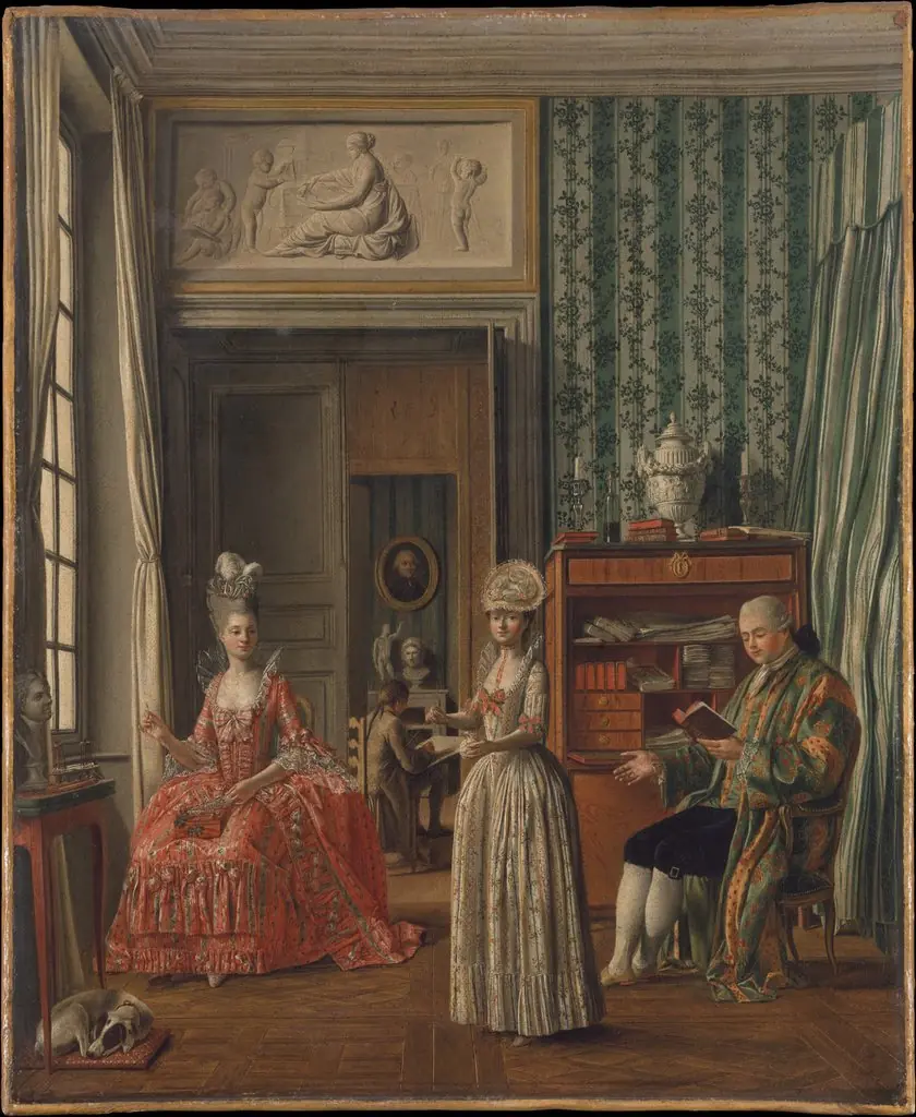 Domestic Scene
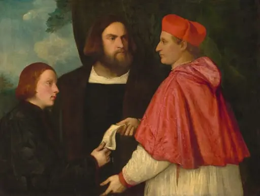 Giacomo and Cardinal Marco, investing Andrea, Abbot of San Zeno, with his Benefice
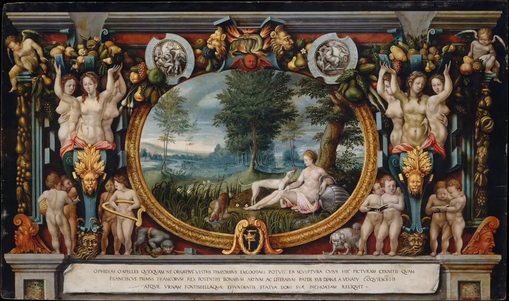 The Nymph of Fontainebleau
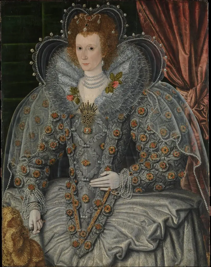 Portrait of a Woman
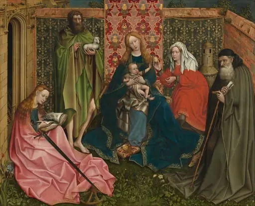 Madonna and Child with Saints in the Enclosed Garden
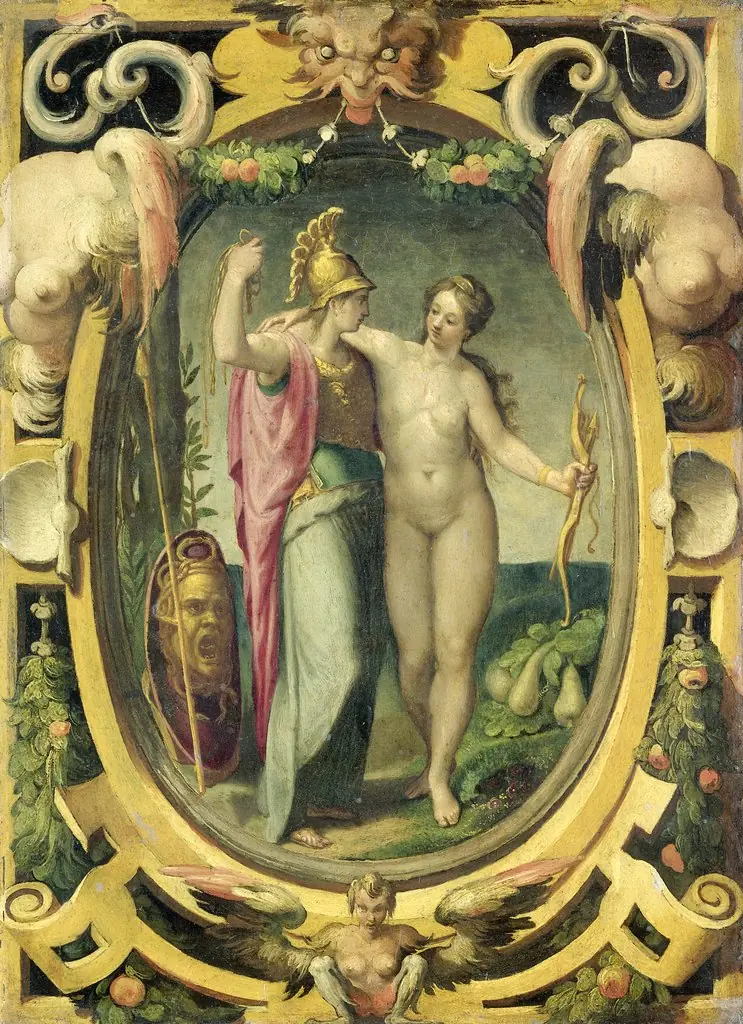 Venus and Minerva
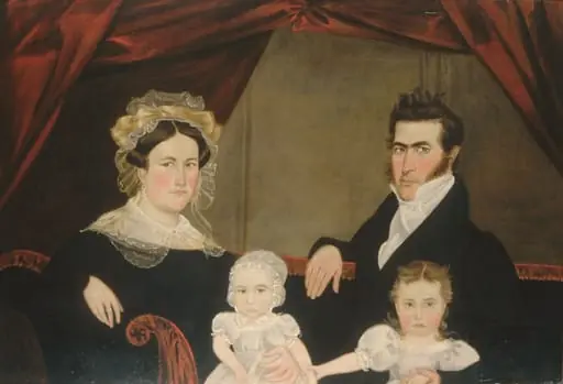 Family Group of Four on Sofa
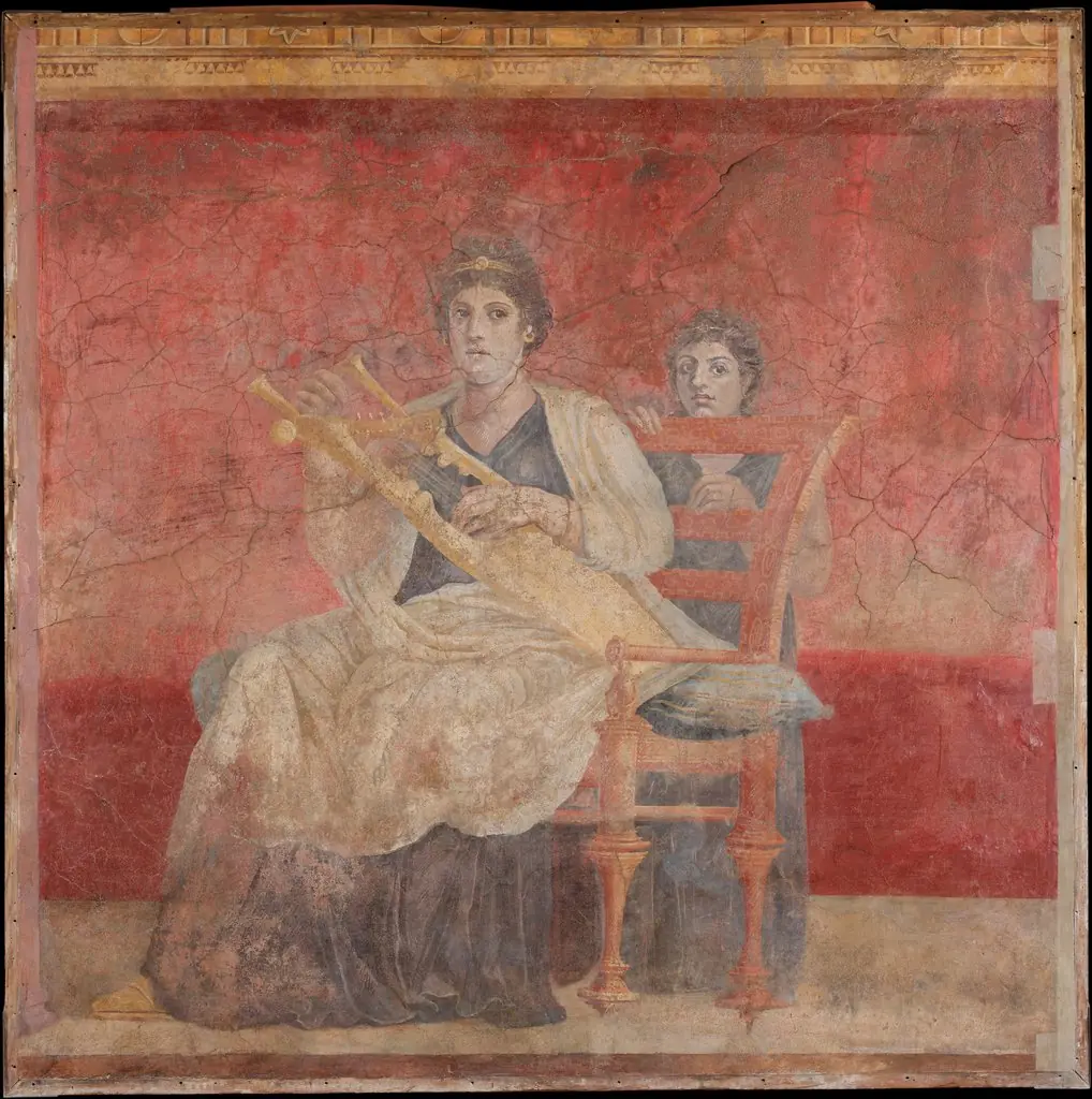 Wall painting from Room H of the Villa of P. Fannius Synistor at Boscoreale