Artwork
Glands of internal secretion
Glands of internal secretion is a drawing by Mol, L. It dates from 1958 and is held in the collection of the Leiden University Libraries. Created in 1958 by L.
About this work
Overview
It is part of the collection at the Museum of Ethnography, where it is preserved as a record of mid-20th-century medical visualization practices.
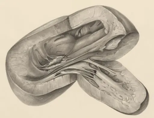
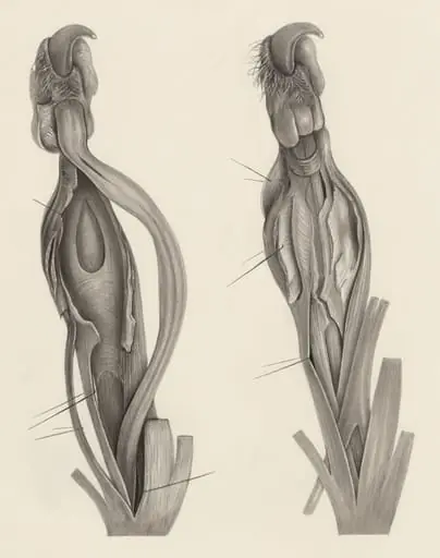
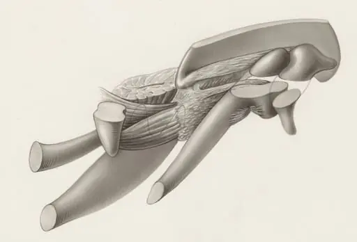
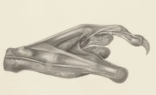
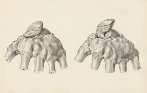
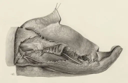
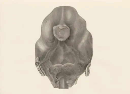
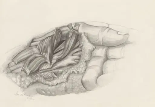
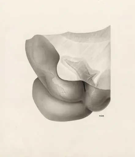
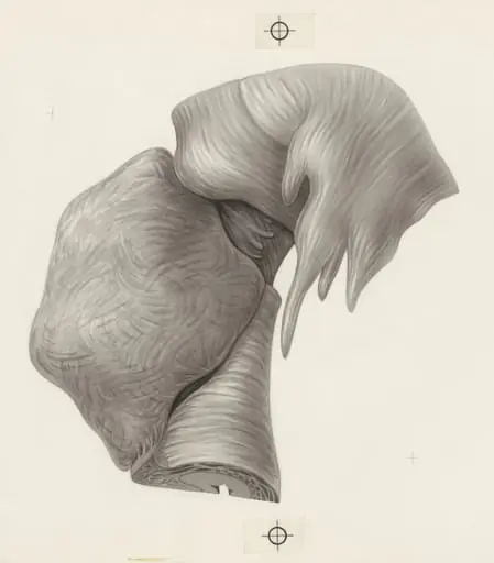
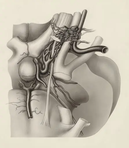
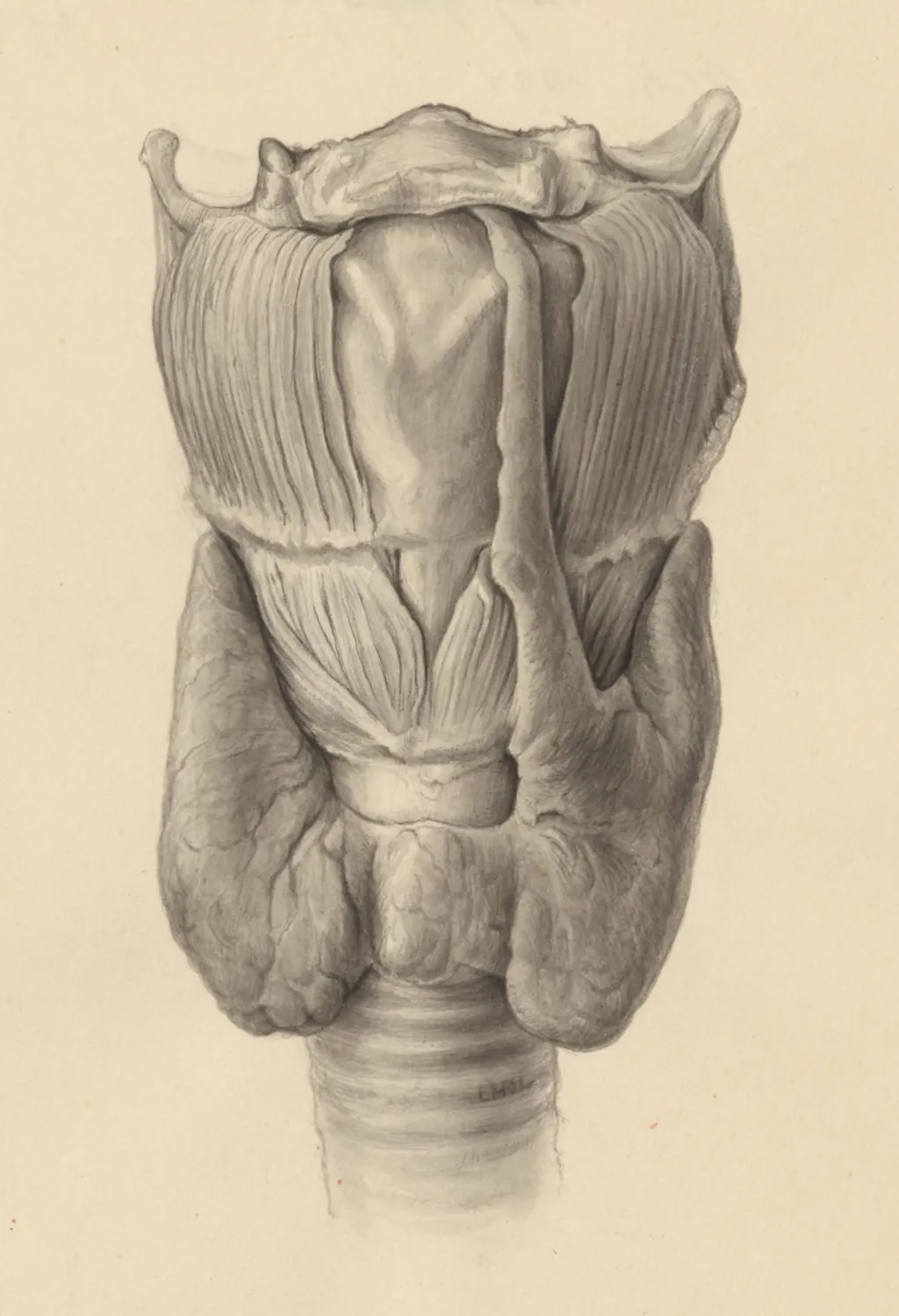
Created in 1958 by L. Mol, this detailed anatomical drawing depicts the human throat and neck region, focusing on internal glands and musculature. Rendered entirely in black and white with meticulous shading, the work serves as a scientific illustration rather than a decorative piece. It is part of the collection at the Museum of Ethnography, where it is preserved as a record of mid-20th-century medical visualization practices.
Subject & Meaning
The drawing isolates the endocrine and muscular structures of the neck, including the thyroid, parathyroid, and associated connective tissues. Its purpose is didactic: to clarify anatomical relationships for educational or professional use. The absence of color and external context emphasizes functional accuracy over aesthetic expression, positioning the image as a tool for understanding human physiology.
Technique & Style
Executed with fine linework and graduated tonal shading, the illustration achieves a high degree of realism. The artist used cross-hatching and stippling to differentiate tissue densities and spatial depth. No outlines dominate; instead, form emerges through subtle gradations of gray, reflecting a commitment to precision over stylistic flourish. The technique aligns with medical drafting traditions of the era.
History & Provenance
The drawing was produced in 1958 by L. Mol, whose full identity remains obscure in public records. It entered the Museum of Ethnography’s collection shortly after creation, likely as part of a broader archive of scientific illustrations. Its preservation suggests institutional recognition of its value as a pedagogical artifact, though its original commission or context is undocumented.
Context
In the late 1950s, anatomical illustration remained a vital medium in medical education, preceding widespread photographic documentation. This work reflects a transitional period where hand-drawn precision still held authority in scientific communication. Similar illustrations were used in textbooks and training manuals, particularly in regions with limited access to advanced imaging technology.
Legacy
Though not widely published or exhibited, the drawing endures as a quiet example of mid-century medical artistry. It contributes to the historical record of how human anatomy was visually interpreted before digital rendering. Its presence in an ethnographic museum underscores the cultural significance of scientific imagery beyond clinical use.
Artist & collection
Artist
This artist carved what most artists skip: the quiet, slippery parts inside us. You’ve seen the glossy posters of muscles and bones, but they focused on the messy, squishy stuff—ducts, glands, half-formed hearts—that…