Artwork
Muscular tissue of the claw of a gray four-eyed opossum
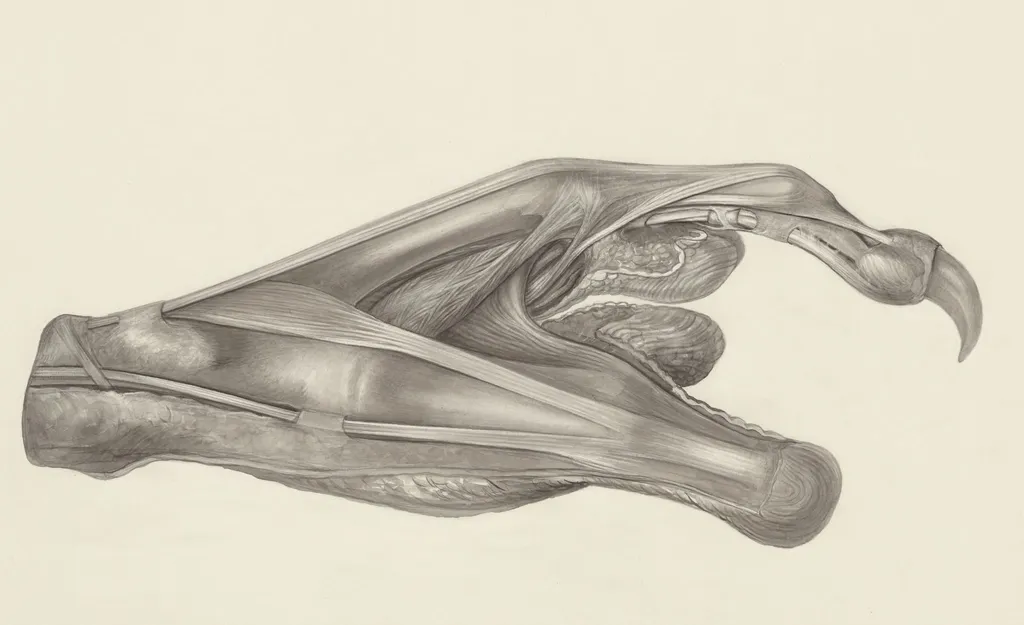
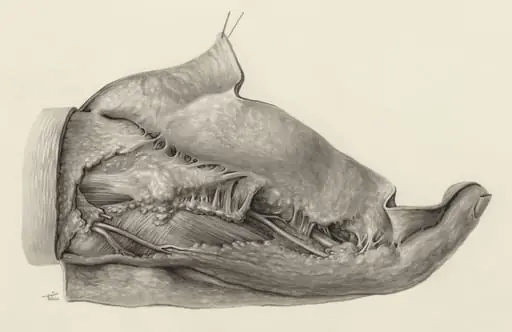
Muscular tissue of the claw of a gray four-eyed opossum is a drawing by H.G, Wetselaar. It dates from 1970 and is held in the collection of the Leiden University Libraries. Created around 1970 by H.
About this work
Overview
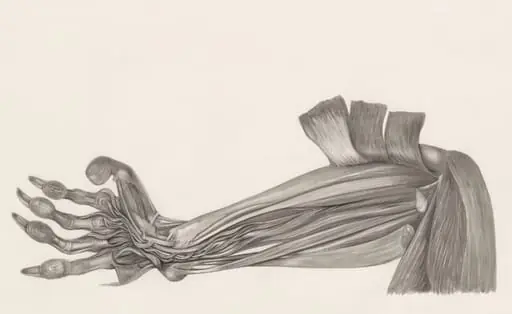
Wetselaar, this detailed anatomical drawing depicts the muscular structure within the claw of a gray four-eyed opossum.
Created around 1970 by H.G. Wetselaar, this detailed anatomical drawing depicts the muscular structure within the claw of a gray four-eyed opossum. Rendered in ink or pencil, the image captures fine biological details with clinical precision. It was produced as part of scientific documentation and is preserved in the collection of the Museum of Ethnography, where it serves as a record of zoological anatomy rather than an artistic statement.
Subject & Meaning
The drawing isolates the claw’s internal musculature, revealing how tendons and muscle fibers attach to and move the bony structure. Rather than portraying the animal as a whole, it focuses on the mechanics of locomotion and grip—key adaptations in arboreal mammals. This level of detail suggests an intent to understand functional morphology, not merely to illustrate appearance.
Technique & Style
The artist employed fine, controlled linework to map the complex interplay of muscle bundles and connective tissue. Shading is minimal; form is defined by contour and cross-hatching that follows anatomical planes. The style resembles scientific illustrations from 19th-century biology texts, prioritizing clarity and accuracy over aesthetic flourish, reflecting a tradition of hand-drawn anatomical study.
History & Provenance
The work was likely produced for academic or research purposes during a period when photographic and radiographic methods were still limited in capturing fine soft-tissue structures. It entered the Museum of Ethnography’s collection as part of a broader archive of zoological studies, possibly linked to field research or comparative anatomy projects in the late 20th century.
Context
Before the widespread use of medical imaging, detailed hand-drawn illustrations were essential tools in biological education and research. Artists like Wetselaar collaborated with scientists to translate microscopic or internal structures into visible, teachable forms. This drawing exemplifies how visual precision supported zoological knowledge in an era reliant on manual observation and rendering.
Legacy
Though largely superseded by digital imaging, such drawings remain valuable for their interpretive clarity and the insight they offer into historical scientific practices. This piece contributes to a legacy of visual documentation that bridged art and biology, preserving anatomical knowledge through meticulous observation before technological alternatives became standard.
Artist & collection
Artist
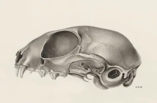
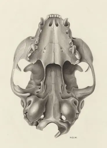
H.G. Wetselaar spent his days hunched over microscopes in a quiet Leiden lab, sketching what most people ignore. His pencil caught the raw architecture of bodies we pretend are smooth—like the knotted muscles of a…

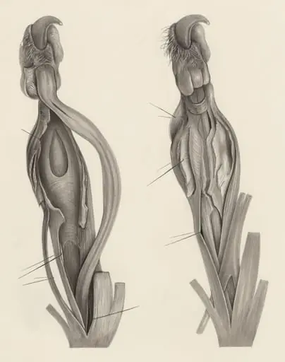

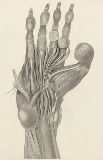
 Muscular tissue of the hand and finger
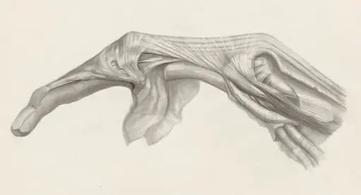 Muscular tissue of the finger
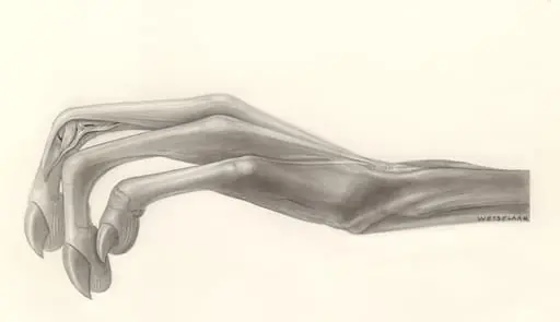
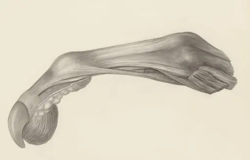 Muscular tissue of the finger of a rhesus macaque
 Muscular tissue of the arm of a rhesus macaque
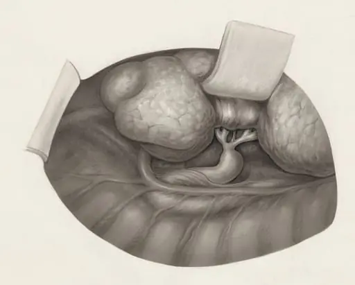
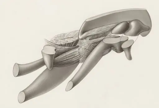 Musculoskeletal system of an embryo
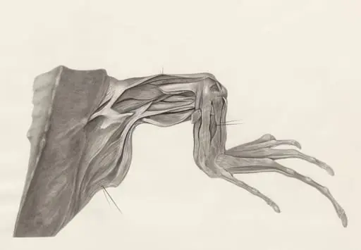 Muscular tissue of a lizards leg and claw
 Muscular tissue of the upper extremities
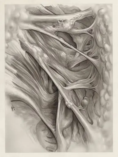 Muscular tissue of the hand or fingers