Artwork
Muscular tissue of a joint
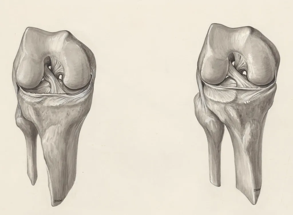
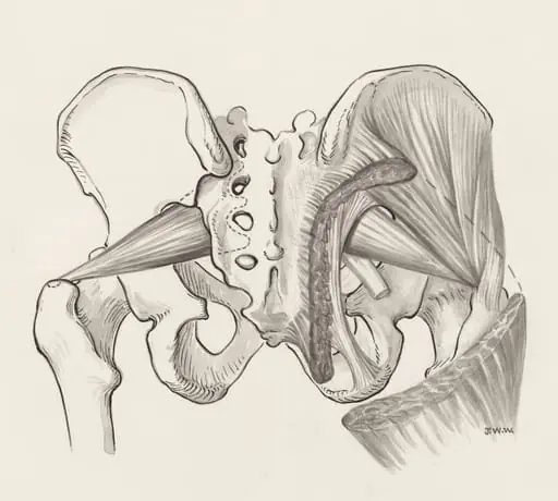
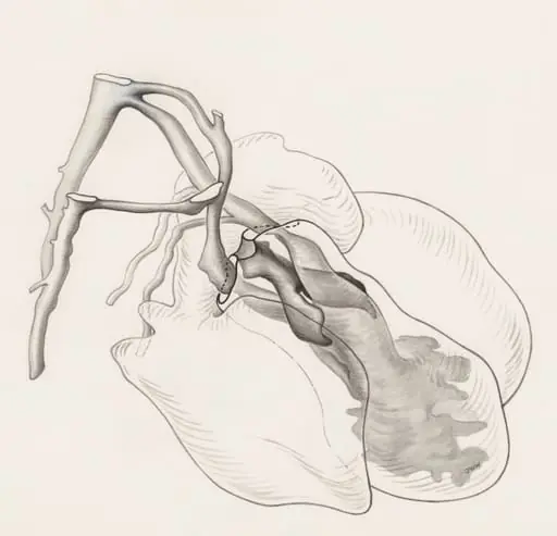
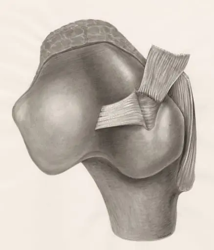
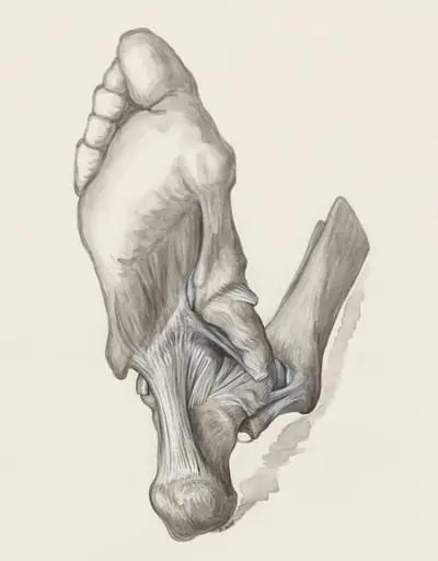
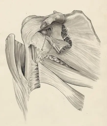
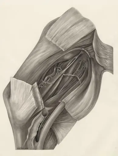
Muscular tissue of a joint is a drawing by J Wetselaar-Whittaker. It dates from 1970 and is held in the collection of the Leiden University Libraries. This drawing depicts the anatomical structure of a human knee joint rendered in monochrome ink.
About this work
Overview
Unlike decorative illustrations, its purpose was pedagogical—designed for medical instruction in the early 19th century.
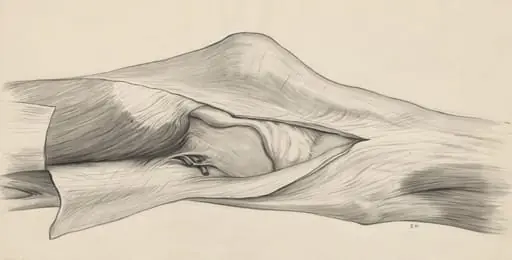
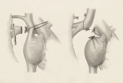
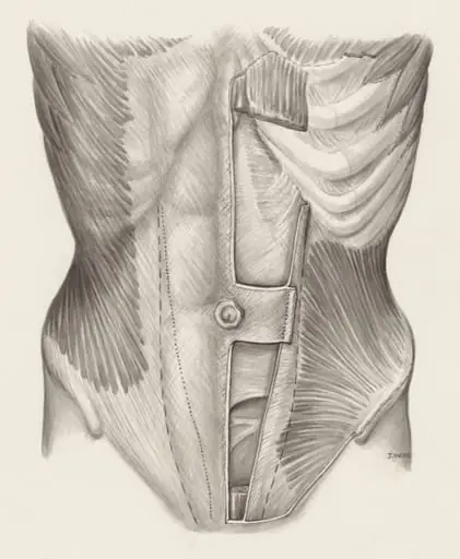
This drawing depicts the anatomical structure of a human knee joint rendered in monochrome ink. Executed with a brush, it employs subtle gray washes and deliberate white highlights to define form. Unlike decorative illustrations, its purpose was pedagogical—designed for medical instruction in the early 19th century. The precision of its lines and tonal gradations reflects a scientific intent rather than artistic flourish.
Subject & Meaning
The drawing isolates the knee’s ligaments, tendons, and bony surfaces with clinical clarity. Each element is labeled implicitly through spatial arrangement and contrast, not text. Its function was to aid surgical trainees in recognizing anatomical relationships under varying light conditions. The absence of skin or muscle tissue focuses attention solely on the joint’s internal architecture, emphasizing structural integrity over aesthetics.
Technique & Style
The artist used a brush to apply diluted gray ink in layered washes, building depth through controlled transparency. White pigment was applied sparingly with a fine tool to accentuate raised surfaces and edges, creating the illusion of volume. The technique avoids heavy outlines, relying instead on tonal transitions to distinguish tissue types. This method mirrors contemporary anatomical atlases that prioritized optical accuracy over ornamentation.
History & Provenance
The work is attributed to the circle of J. Wetselaar-Whittaker, a 19th-century medical illustrator active in European anatomical schools. Likely produced for use in surgical training programs, it belongs to a broader tradition of hand-drawn anatomical plates circulated among medical institutions. Its survival suggests it was valued as a reference tool, possibly used in classrooms or private collections of practicing surgeons.
Context
In the early 1800s, surgical education relied heavily on visual aids due to limited access to cadavers and rudimentary printing. Drawings like this filled a critical gap, offering detailed, reproducible models of anatomy. Similar works were produced in Paris, Edinburgh, and Vienna, often by artists trained in both art and medicine. This piece reflects the growing professionalization of surgery and the demand for standardized visual instruction.
Legacy
Though overshadowed by later photographic and printed anatomical texts, such hand-drawn illustrations laid the groundwork for modern medical visualization. Their emphasis on clarity, proportion, and structural accuracy influenced generations of anatomical illustrators. Today, they are studied as historical artifacts that reveal how medical knowledge was transmitted visually before the advent of digital imaging.
Artist & collection
Artist
This artist made precise studies of the body—drawings and sculptures that cut straight to the bones, muscles, and tissues.