Artwork
Muscular tissue of a lizards leg
Muscular tissue of a lizards leg is a drawing by H.G, Wetselaar. It dates from 1970 and is held in the collection of the Leiden University Libraries. This drawing, created around 1970 by H.
About this work
The level of detail in the drawing suggests that it may have been created for educational or scientific purposes.
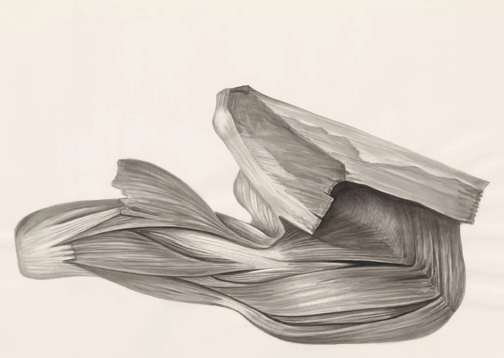
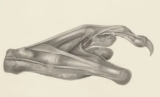
This drawing depicts the muscular tissue of a lizard's leg. The artist has rendered the muscles in a detailed, realistic manner, with visible striations and texture. The drawing is done in a range of grays, with darker shading used to create depth and dimension.
The level of detail in the drawing suggests that it may have been created for educational or scientific purposes. The artist's use of cross-hatching and stippling techniques adds to the overall sense of realism and texture.
If you're interested in learning more about the artist behind this piece, you can look up Wetselaar, H.G, (1926-).
Overview
This drawing, created around 1970 by H.G. Wetselaar, presents a precise anatomical study of a lizard’s hind limb musculature. Rendered entirely in grayscale, the work emphasizes structural clarity over aesthetic flourish. It was produced for scientific documentation and is part of the collection at the Museum of Ethnography, where it serves as a record of biological observation rather than artistic expression.
Subject & Meaning
The subject is the muscular architecture of a lizard’s leg, depicted with attention to fiber orientation, insertion points, and tissue layering. The focus on anatomical accuracy suggests an intent to aid in comparative biology or zoological education. There is no symbolic or narrative layer; the image functions as a visual reference, capturing the mechanical efficiency of reptilian locomotion through form alone.
Technique & Style
Wetselaar employed fine-line cross-hatching and stippling to simulate the striated texture of skeletal muscle. Variations in tone, achieved through dense line work and graduated shading, convey depth without color. The absence of outlines and the meticulous rendering of individual muscle bundles reflect a draftsmanship aligned with scientific illustration traditions, prioritizing fidelity over stylistic flair.
History & Provenance
The drawing was made by H.G. Wetselaar, a Dutch artist and illustrator active from the mid-20th century, known for biological and ethnographic documentation. It entered the Museum of Ethnography’s collection in the 1970s, likely as part of a broader archive of zoological studies. Its origin as a working illustration, rather than a finished artwork, informs its preservation context within a research-oriented institution.
Context
Created during a period when hand-drawn anatomical illustrations remained vital in biological sciences, this work reflects a pre-digital era of scientific record-keeping. Similar drawings were used in textbooks, field guides, and museum archives to communicate complex anatomical relationships. Wetselaar’s contribution aligns with a tradition of illustrators supporting academic disciplines through visual precision.
Legacy
Though not widely exhibited, the drawing endures as a representative example of mid-20th-century scientific illustration. It contributes to the Museum of Ethnography’s holdings of observational materials, offering insight into how biological knowledge was visually transmitted before digital imaging. Its continued presence underscores the value of manual draftsmanship in the history of natural science.
Artist & collection
Artist
H.G. Wetselaar spent his days hunched over microscopes in a quiet Leiden lab, sketching what most people ignore. His pencil caught the raw architecture of bodies we pretend are smooth—like the knotted muscles of a…

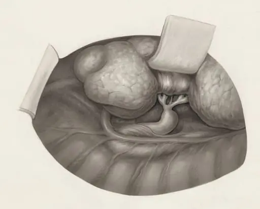
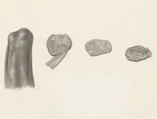 Cross sections of bone and muscular tissue on the hind leg of a lizard
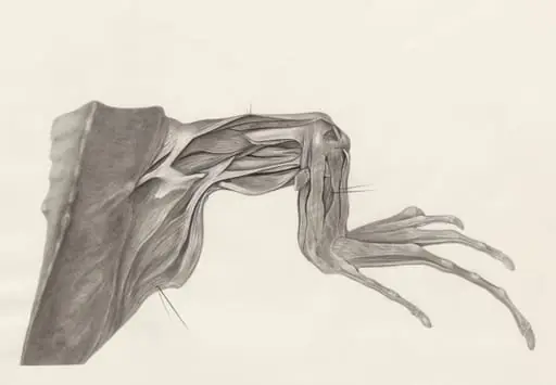 Muscular tissue of a lizards leg and claw
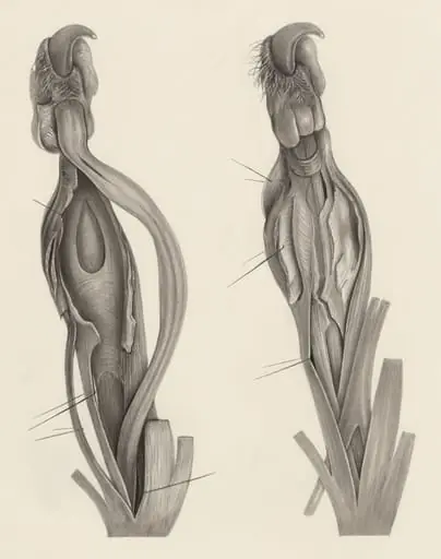
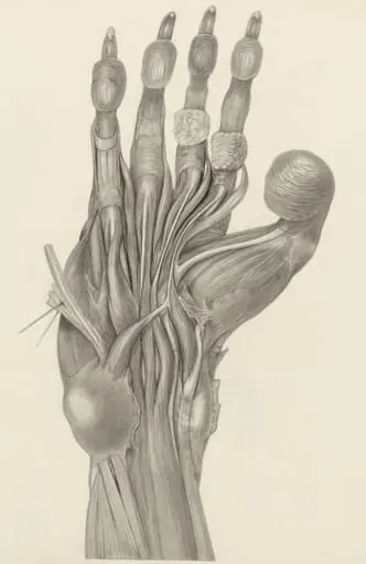
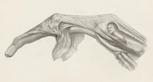 Muscular tissue of the finger
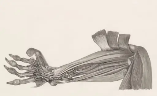
 Muscular tissue of the arm of a rhesus macaque
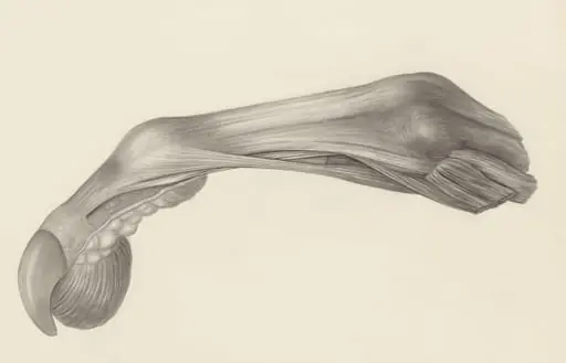 Muscular tissue of the finger of a rhesus macaque
 Muscular tissue of the upper extremities
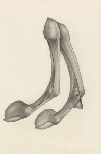 Muscular tissue of a mammal's finger
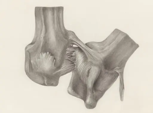 Muscular tissue of a mammal's upper extremities
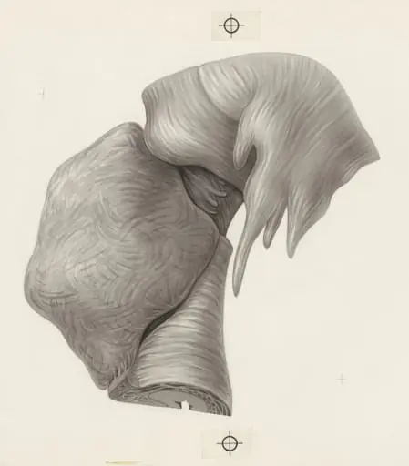 Human tissue