Artwork
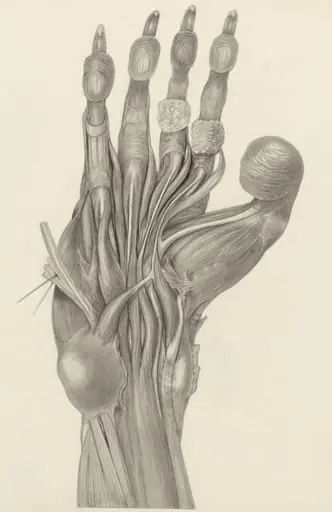
Muscular tissue of the finger of a rhesus macaque
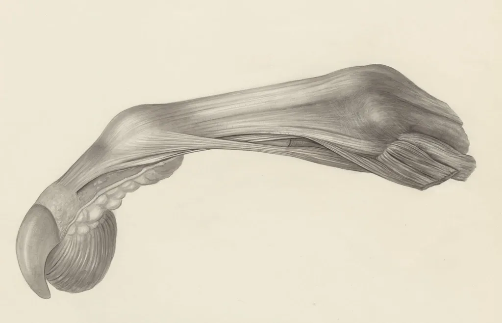
Muscular tissue of the finger of a rhesus macaque is a drawing by H.G, Wetselaar. It dates from 1970 and is held in the collection of the Leiden University Libraries. This detailed anatomical drawing, created around 1970 by H.
About this work
Overview
Rendered in ink and shading, it presents the finger with its skin removed to expose underlying musculature, tendons, and bone.
This detailed anatomical drawing, created around 1970 by H.G. Wetselaar, depicts the internal structures of a rhesus macaque’s finger. Rendered in ink and shading, it presents the finger with its skin removed to expose underlying musculature, tendons, and bone. The work was produced for scientific study and is part of the Museum of Ethnography’s collection, reflecting mid-20th-century practices in biological illustration.
Subject & Meaning
The subject is the precise arrangement of soft and hard tissues in a primate digit. By stripping away the skin, the drawing reveals how muscles originate from bone, transition into tendons, and insert at the joint. This level of anatomical clarity served educational purposes, helping researchers and students understand the mechanical relationships that enable fine motor control in primates.
Technique & Style
The artist employed fine, controlled lines and graded shading to suggest volume and depth without color. Smooth contours define muscle bundles, while subtle tonal transitions indicate curvature and spatial relationships. Cross-hatching and stippling are used sparingly to enhance the three-dimensionality of tendons and connective tissue, emphasizing structural accuracy over aesthetic flourish.
History & Provenance
Created in the early 1970s, the drawing was likely produced as part of a research or teaching project in primatology or comparative anatomy. It entered the collection of the Museum of Ethnography, where it remains as a specimen of scientific visualization. Its preservation underscores the historical value of hand-drawn anatomical records before the dominance of photographic and digital methods.
Context
During the 1960s and 1970s, detailed anatomical illustrations remained essential in biological sciences, particularly when studying non-human primates. Such drawings were often made from dissections, serving as both documentation and pedagogical tools. This piece reflects a tradition of meticulous observation, where accuracy in representation was prioritized over artistic expression.
Legacy
Though largely superseded by imaging technologies, this drawing exemplifies the enduring role of hand-drawn anatomy in scientific communication. It preserves a moment when close observation and manual rendering were the primary means of understanding biological form. Today, it stands as a quiet testament to the discipline and precision required in pre-digital biological research.
Artist & collection
Artist
H.G. Wetselaar spent his days hunched over microscopes in a quiet Leiden lab, sketching what most people ignore. His pencil caught the raw architecture of bodies we pretend are smooth—like the knotted muscles of a…
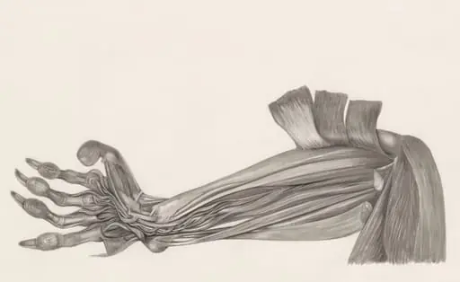

 Muscular tissue of the arm of a rhesus macaque
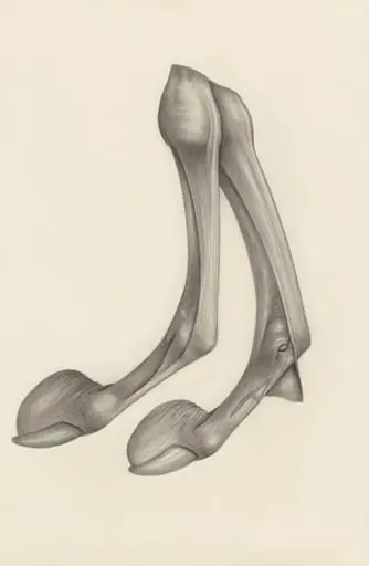 Muscular tissue of a mammal's finger
 Muscular and bone tissue of the hand or fingers
 Muscular tissue of the hand and finger
 Muscular tissue of a mammal's upper extremities
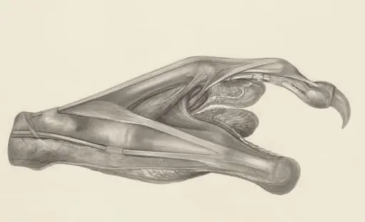
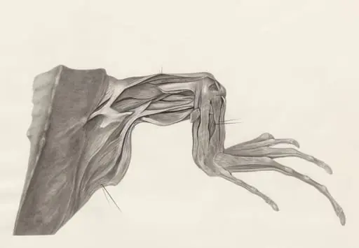 Muscular tissue of a lizards leg and claw
 Muscular tissue of the upper extremities
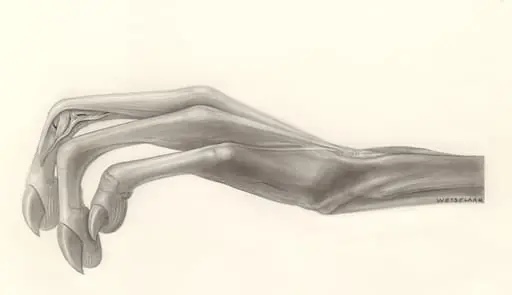 Muscular tissue of the hand