Artwork
Deformed fetal skull, with part of the spinal cord
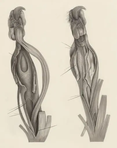
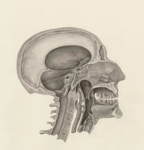
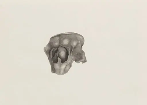
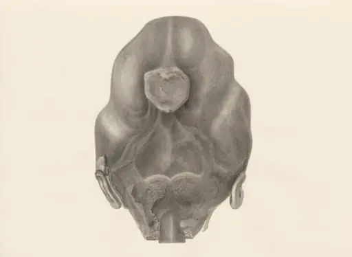
Deformed fetal skull, with part of the spinal cord is a drawing by H.G, Wetselaar. It dates from 1970 and is held in the collection of the Leiden University Libraries. This anatomical drawing, attributed to H.
About this work
Overview
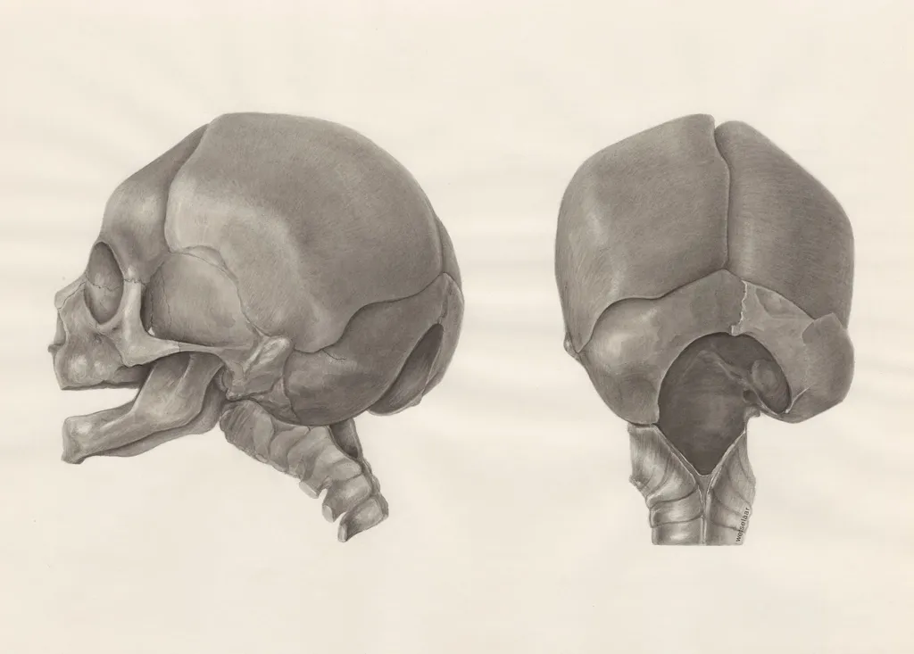
Rendered in precise draftsmanship, it presents two angled views—front-left and back-right—to illustrate structural anomalies.
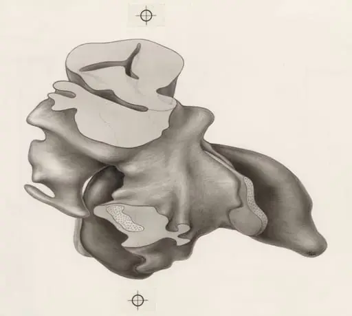
This anatomical drawing, attributed to H.G. Wetselaar and dated circa 1970, depicts a fetal skull with an associated segment of the spinal cord. Rendered in precise draftsmanship, it presents two angled views—front-left and back-right—to illustrate structural anomalies. The work was produced for scientific documentation and is held in the collection of the Museum of Ethnography, where it serves as a record of developmental variation.
Subject & Meaning
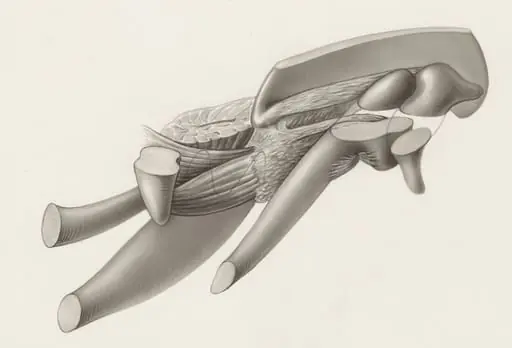
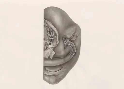
The subject is a malformed fetal skull, likely illustrating a congenital condition affecting cranial development. The inclusion of the upper spinal cord suggests an interest in the relationship between neural and skeletal structures. The open jaw and irregular bone contours indicate pathological change, not idealized anatomy. The image functions as a clinical observation, prioritizing accuracy over aesthetic presentation.
Technique & Style
The drawing employs fine, controlled lines and cross-hatching to model volume and texture. Shading is built through layered strokes, giving the bones a sculpted, three-dimensional presence. The absence of color or background emphasizes structural detail. The hand-drawn quality reflects meticulous observation, typical of anatomical illustrations used in medical education and research during the mid-20th century.
History & Provenance
Created around 1970, the drawing entered the Museum of Ethnography’s collection as part of a broader anthropological or medical archive. Its origin may be tied to a research project examining fetal development or congenital anomalies in specific populations. While the artist’s full professional context remains unclear, the work aligns with mid-century practices of documenting biological variation for scholarly use.
Context
In the 1970s, anatomical illustration remained a vital tool in medical anthropology and embryology, especially where photography was limited or insufficient for capturing fine structural detail. This drawing reflects a period when hand-drawn records were still standard in clinical and ethnographic studies, bridging scientific rigor with manual skill to preserve rare biological specimens.
Legacy
The drawing endures as a quiet example of pre-digital anatomical documentation. It contributes to historical records of how medical professionals observed and represented developmental anomalies before widespread imaging technologies. Its preservation in an ethnographic museum underscores its value not only as a medical artifact but as a cultural record of scientific practice.
Artist & collection
Artist
H.G. Wetselaar spent his days hunched over microscopes in a quiet Leiden lab, sketching what most people ignore. His pencil caught the raw architecture of bodies we pretend are smooth—like the knotted muscles of a…